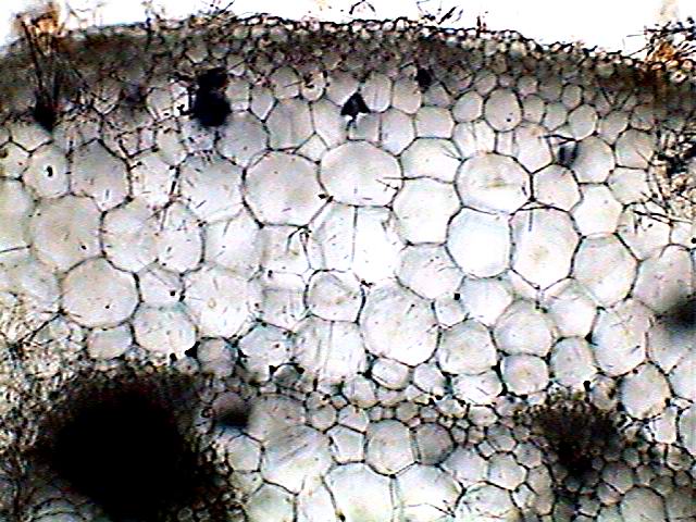
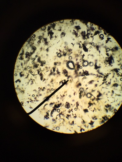
Monocot Root Under Microscope 10x
Sharp razor brush dropper needles watch glass microscopic slides cover slips safrannin glycerine and compound microscope.

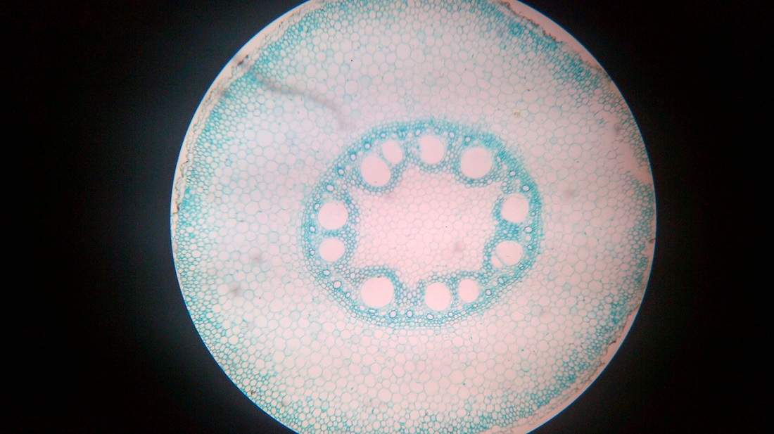
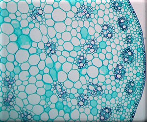
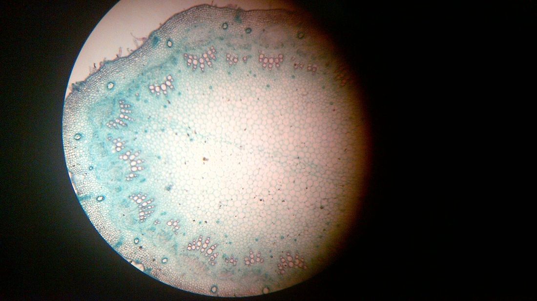
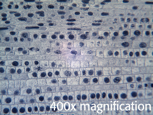
Monocot root under microscope 10x. Saved from vcbio. Both monocot and dicot roots belong to plants. Telophase the final phase of cell division will appear as two nuclei are formed and have little or no cell wall between.
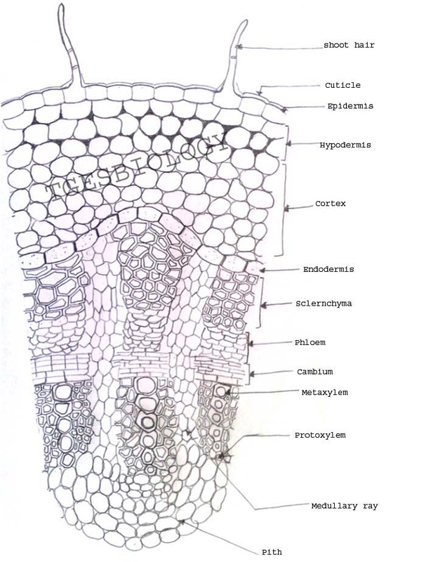
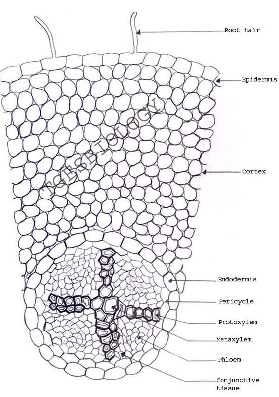
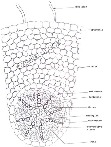
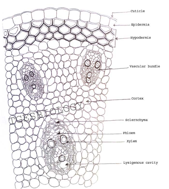
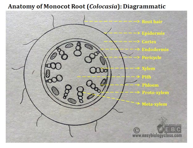
Roots mounted together for comparison. O anatomically the monocot root has been differentiated into the following parts. Transverse section took through the internode of the stem.
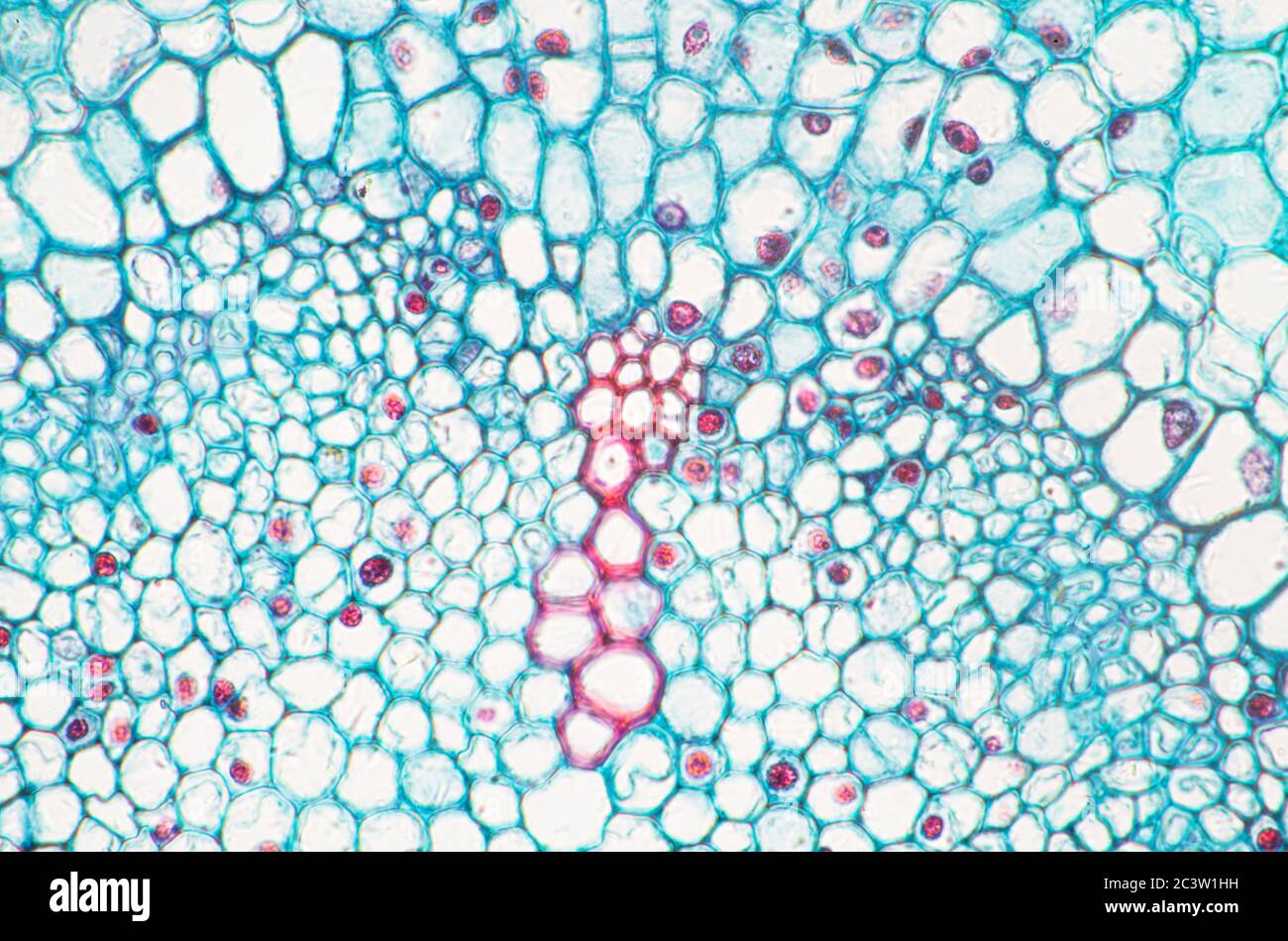
Locate the vascular cylinder and switch to high power. Take 2 3cm long pieces of the material. Identify the xylem and phloem cells and note their locations.
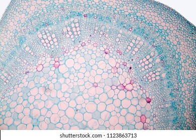
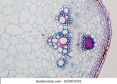
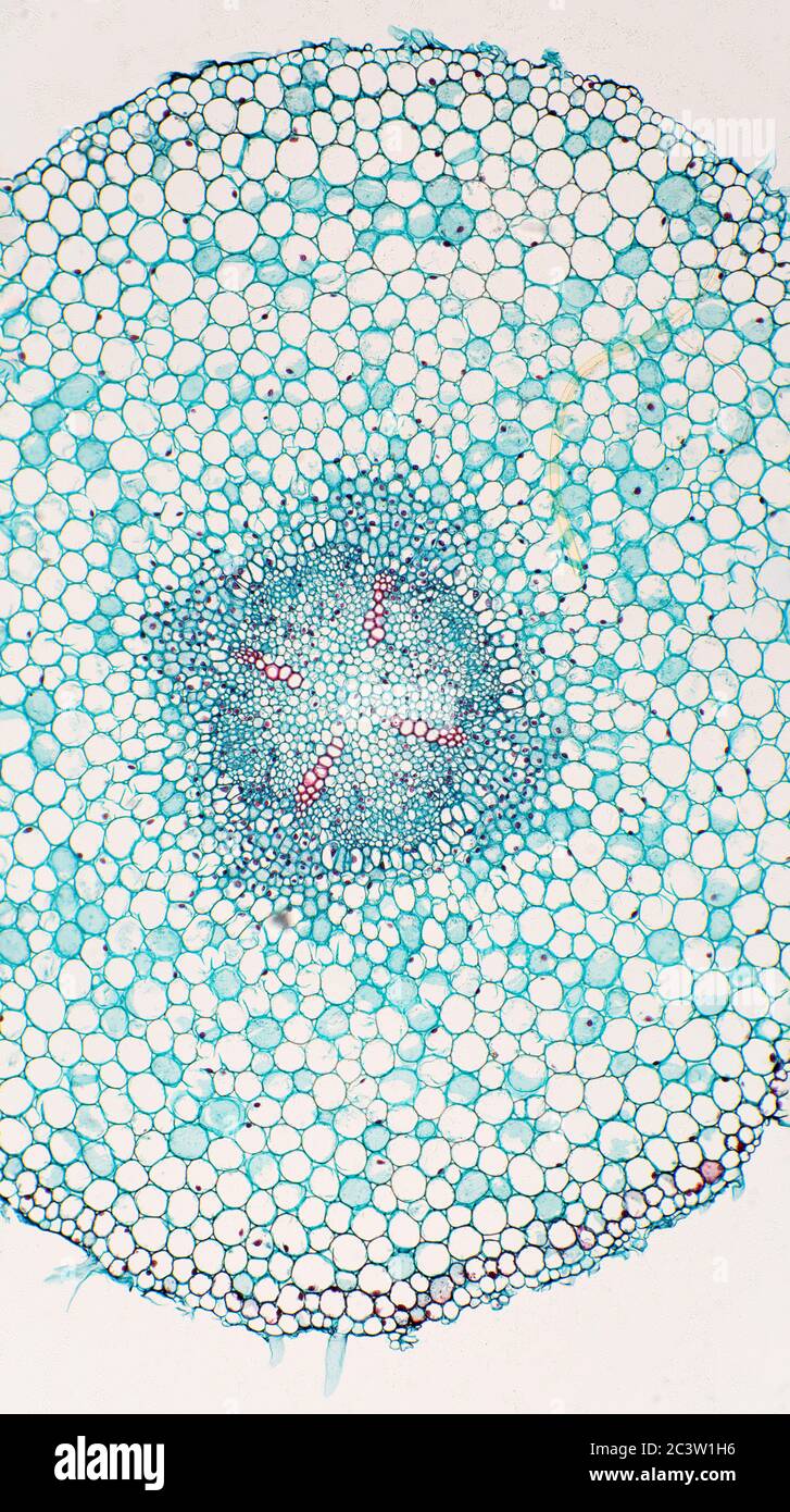
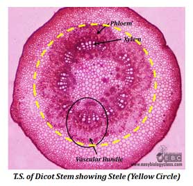
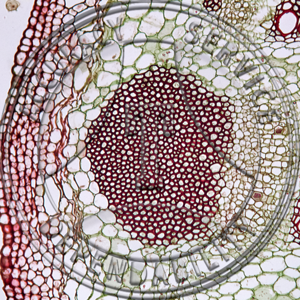
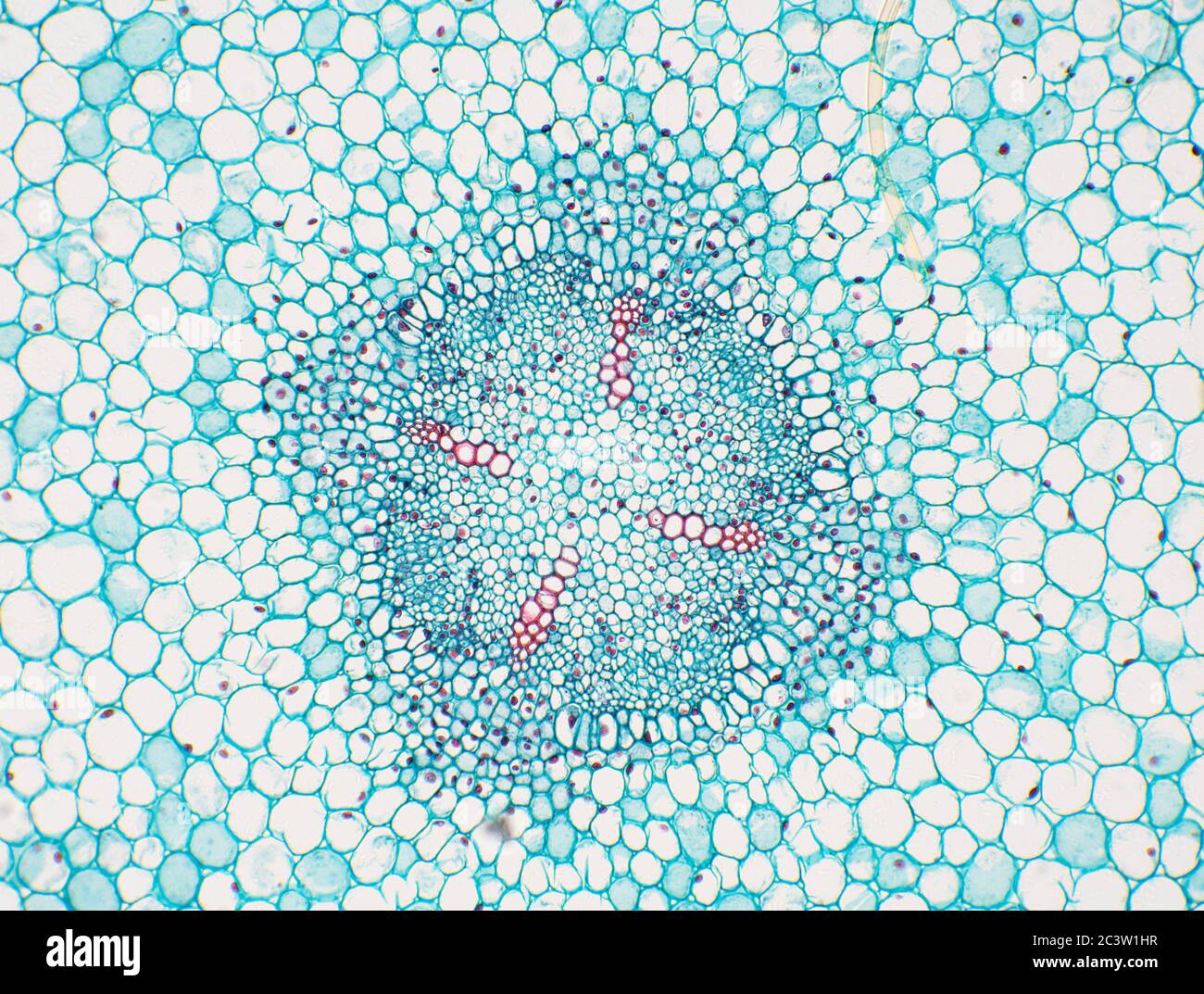
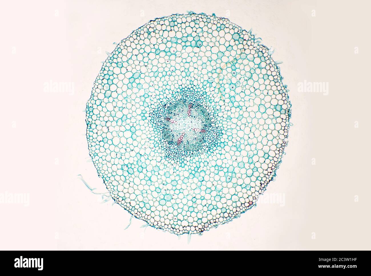
O anatomically the dicot stem has the following regions. Jan 24 2017 monocot root cell transverse section through a monocot root. Root cross section of a monocot plant zea mays maize corn.
Using a microscope its possible to view and identify these cells and how they are arranged epidermal cells spongy cells etc. To prepare temporary stained glycerine mounts of transverse sections of stem and root of dicot and monocot plants. Monocot and dicot differ from each other in four structures.
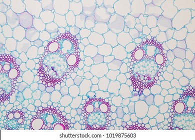
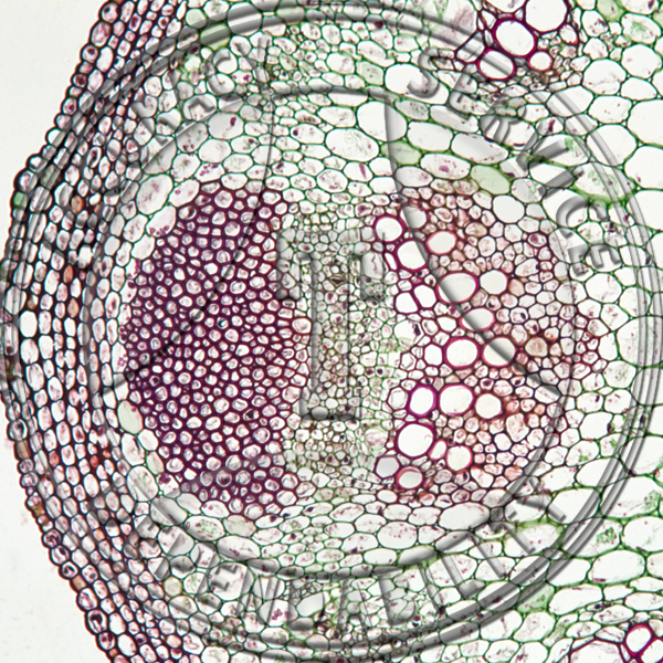
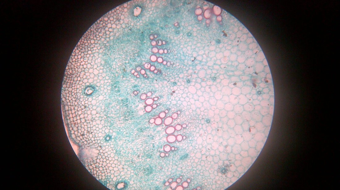
While monocot root contains xylem and phloem in another manner forming a circle. This is another panorama photomicrograph assembled from four individual images. 30 1898 shows corn zea and buttercup ranunculus.
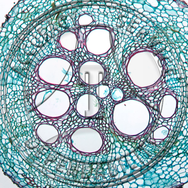
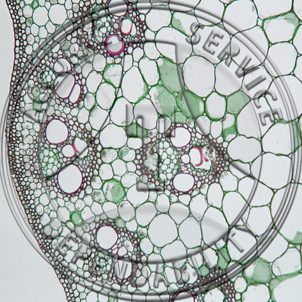
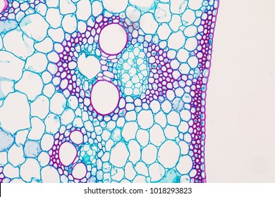
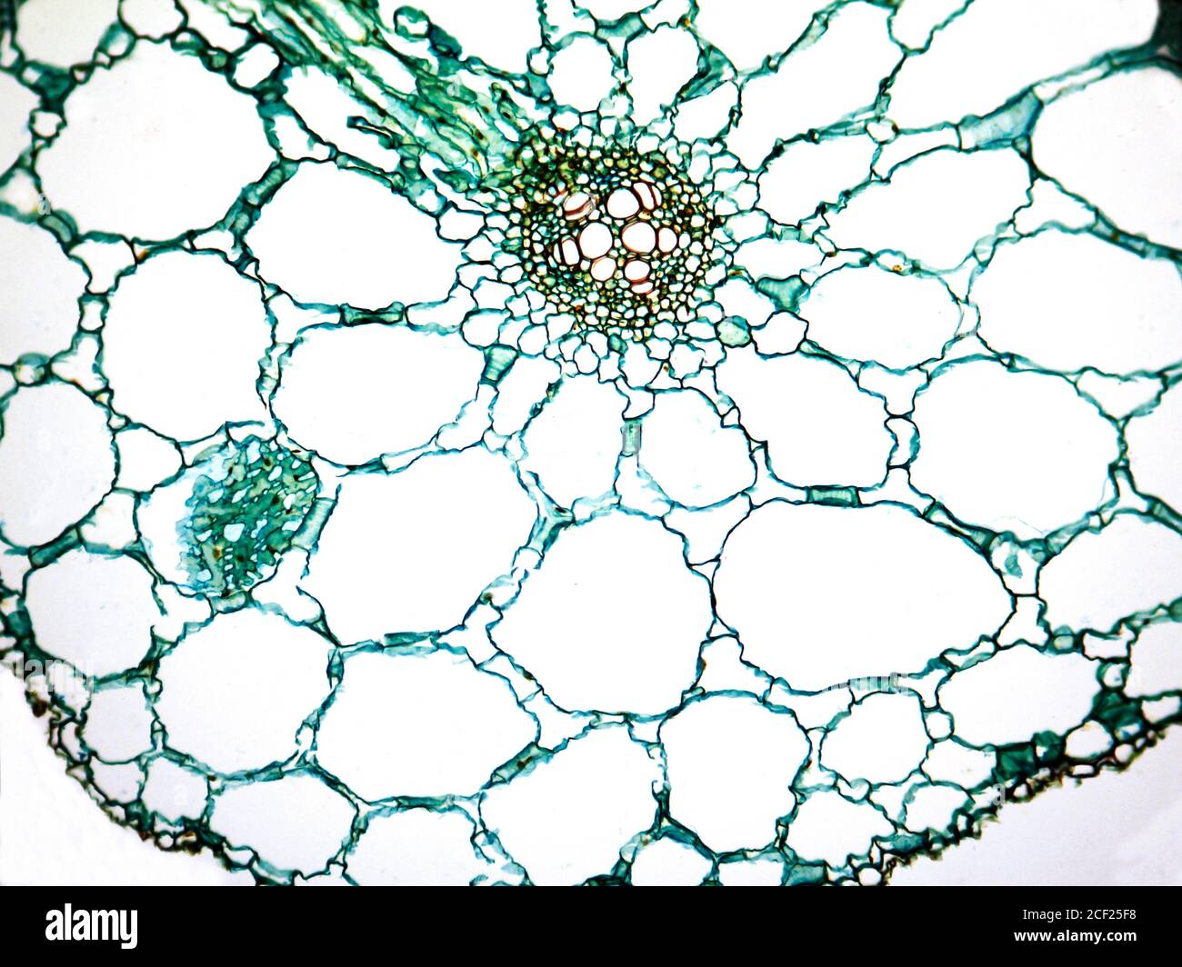
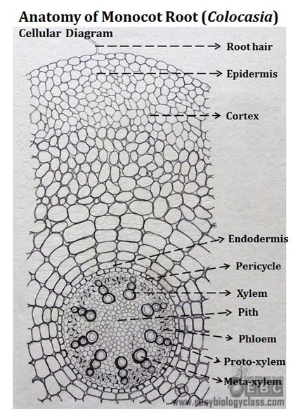
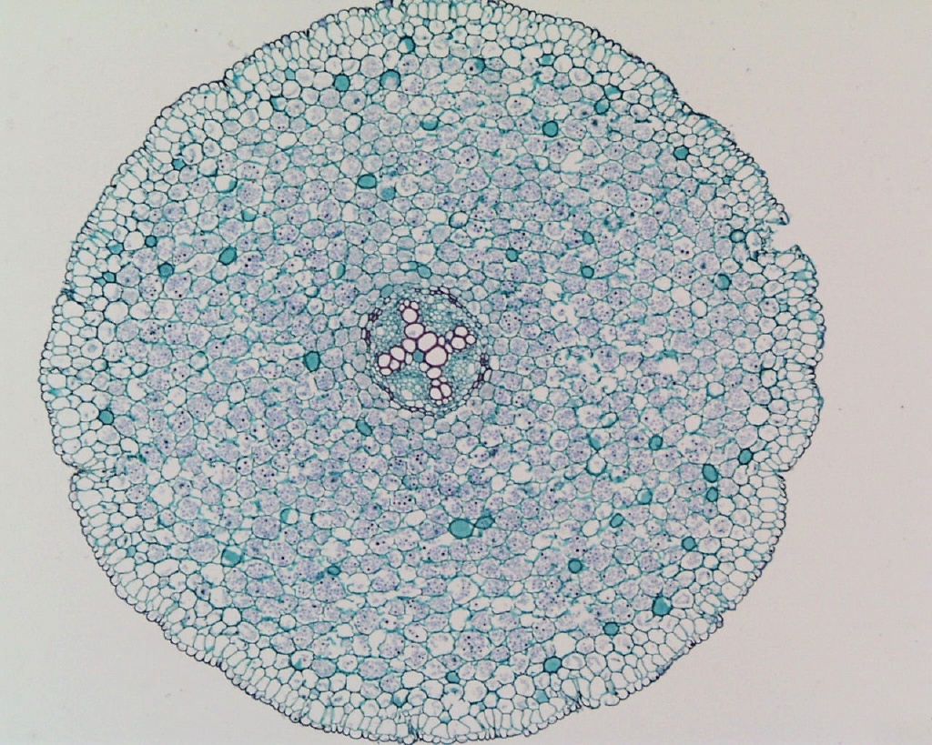
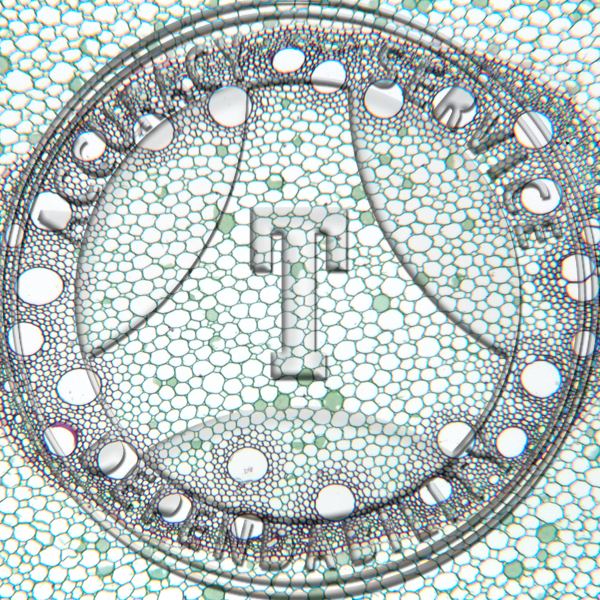
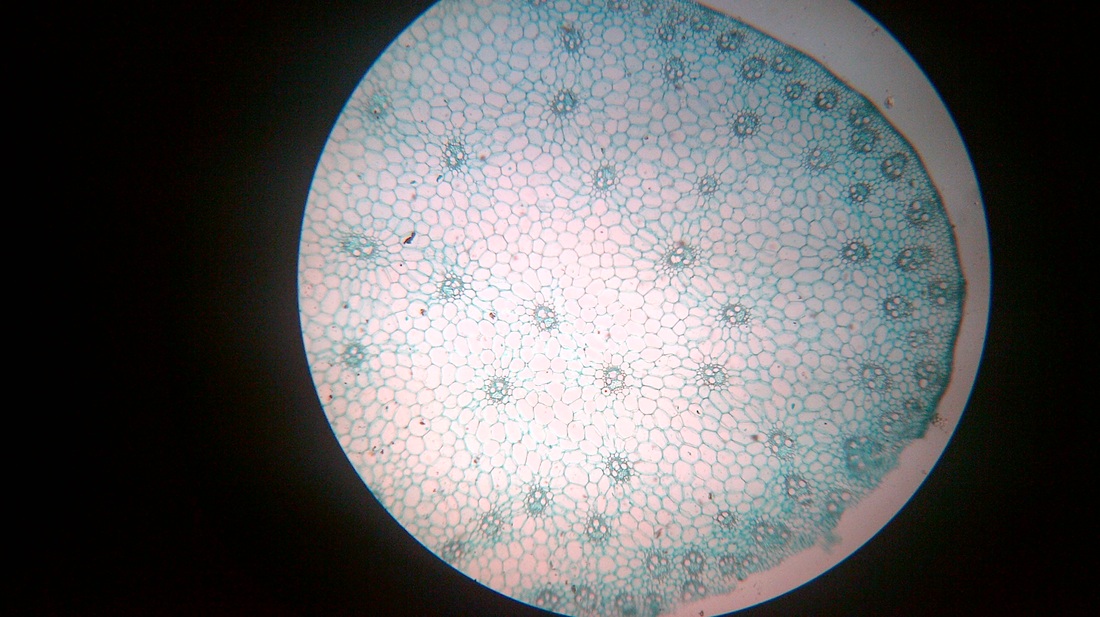
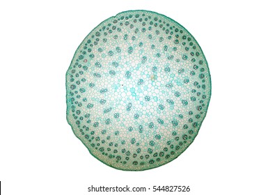
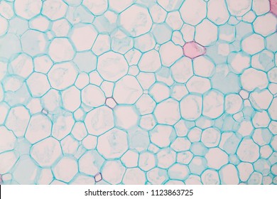
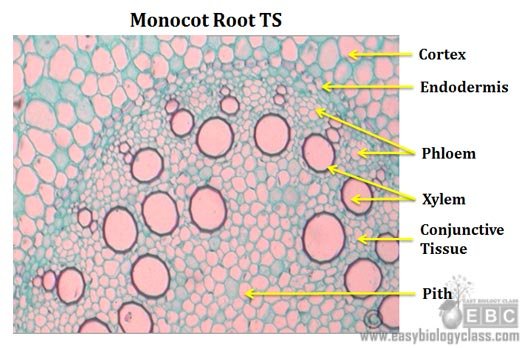
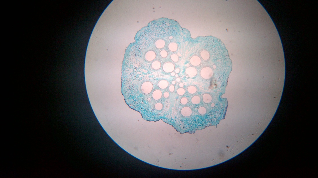
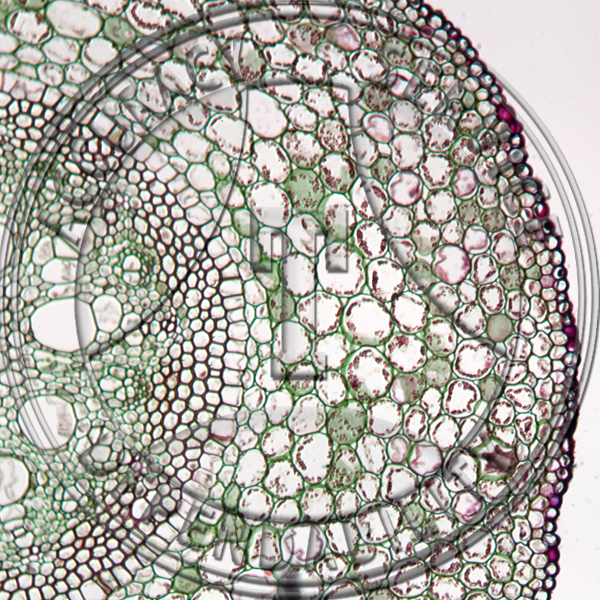
The image shows a cross section of zea mays maize a monocotyledonous plant monocotthe picture shows epidermis the outside layer of cells endodermis inside ring shaped layer of smaller cells and vascular tissue the larger cells inside. Anatomy of monocot root monocot root cross section under microscope with diagram o the anatomical features of a monocot root can be studied through a cross section cs through the root. How is the arrangement of the vascular cylinder different.
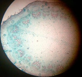
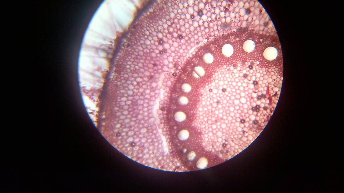
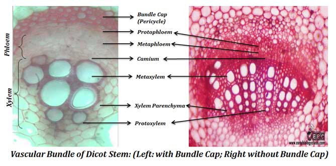
O the components of cortex and stele are together known as ground tissue. O the anatomy of dicot stem is studied by a ts. The monocot roots are fibrous while that of dicot is.
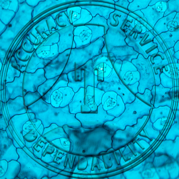
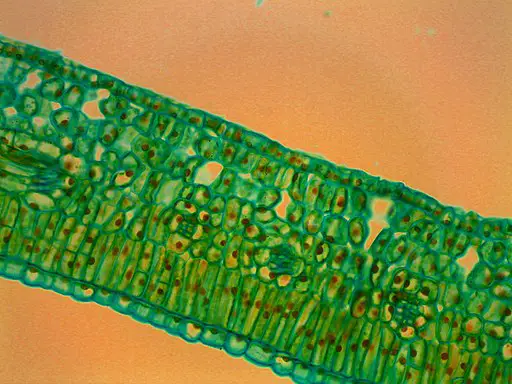
Using the microscope view the monocot root slide under low power. Leaves stems roots and flowersthe difference between dicot and monocot root is dicot root contains xylem in the middle and phloem surrounding it. Viewing the leaf under the microscope shows different types of cells that serve various functions.
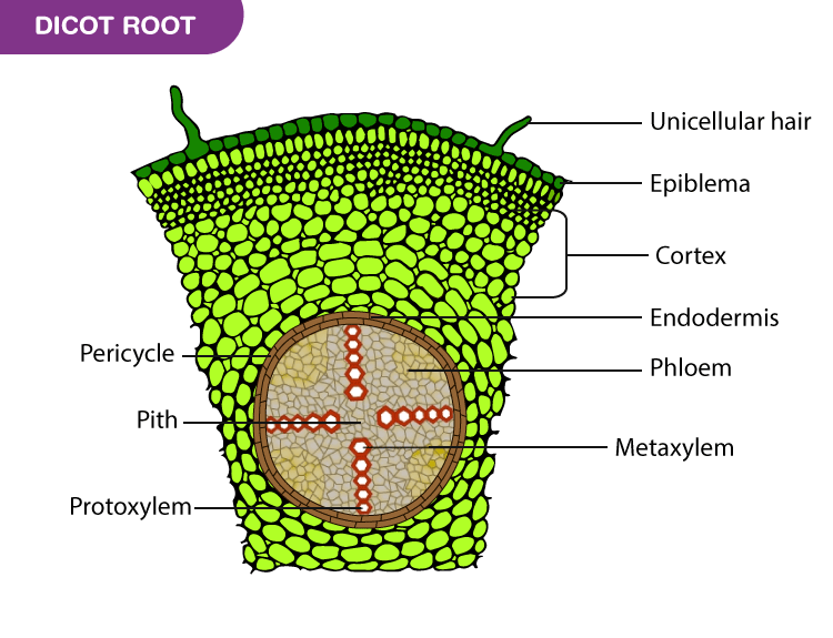
Now look at the dicot root. Biology art marine biology microscopic photography macro photography microscopic images things under a microscope nature drawing zoology science art. 30 1892 shows carrion flower smilax and buttercup ranunculus.
Prepared slide of monocot and dicot roots.


